PET
What is Malignant Mesothelioma Cancer?
Malignant mesothelioma cancer is a rare medical condition that impacts the mesothelial cells of the serous membranes. The most common type of this cancer is pleural mesothelioma, which disrupts the lining of the lungs. Each year, approximately 2,000-3,000 cases of mesothelioma cancer are diagnosed.
Malignant mesothelioma cancer disturbs the r lining of certain large cavities in the body. These cavities, labeled the serous cavities, serve as a shield for a number of vital organs in the body, including the lungs. The membranes that border these cavities protect several organs from abrasions and friction that occurs as the organs move rub each other during typical movement, such as breathing. Said membranes derive from mesothelial cells, which form to create the mesothelium—the tissue layer of the serous membranes.
Malignant mesothelioma is observed in three distinct forms:
Pericardial Mesothelioma Cancer: Forms in the pericardium (lining of tissue surrounding the heart).
Peritoneal Mesothelioma Cancer: Forms in the peritoneum (membrane surrounding the abdomen). A subset of this type of mesothelioma may also form in the male testicles, specifically the peritoneum--the lining surrounding the scrotum.
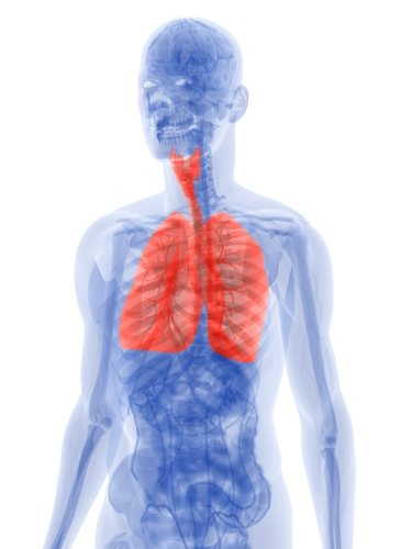
Pleural Mesothelioma Caner: As previously stated, pleural mesothelioma cancer is the most common form of mesothelioma. Pleural mesothelioma cancer disrupts the lining of the lungs.
All types of mesothelioma derive from a localized tumor. The tumor will lie dormant for several years; however, proliferation is rapid--the disease will quickly spread to surrounding organs and tissue. The majority of mesothelioma patients contract the disease from previous exposure to asbestos filaments. It must be noted; however, that not every individual who is in contact with asbestos-containing materials will develop mesothelioma cancer.
Although the medical condition is abnormally rare, it is extremely deadly. When finally detected, mesothelioma cancer is often in its advanced stages. As a result, prognosis for mesothelioma patients is deemed far bleaker than patients with other types of cancer that are frequently detected earlier. On average, survival rates following a mesothelioma diagnosis is only 1 to 2 years. This timeframe; however, fluctuates based on the type of mesothelioma cancer found.
Malignant mesothelioma cancer is difficult to accurately diagnose because the symptoms are slow-developing—mesothelioma patients will not notice symptoms for 10-15 years following infection. Furthermore, mesothelioma cancer, at its roots, possesses similar cellular makeups to other forms of cancer. These characteristics make the disease nearly impossible to detect; however, with advancements in technology and innovations in diagnostic tests, the early-stage diagnosis of mesothelioma cancer has improved greatly. A PET or Positron Emission Tomography machine is an example of the strides we have made in detecting this horrible disease.
What is a PET Scan?
A PET scan (short for Positron Emission Tomography) is a nuclear imaging technique that produces a three-dimensional image of body processes. The PET machine detects pairs of gamma rays that are emitted indirectly through a positron-emitting tracer. These rays are introduced to the patient on a biologically active molecule. Following this introduction, three-dimensional images of tracer concentration within the patient’s body are constructed by computer analysis.
PET scanning provides more detailed information than other imaging devices, such as MRI or CT scans. A PET image will detect biological alterations to reveal extremely small tumors within the body. Able to detect whether these tumors are benign or malignant, a PET scan also reveals the extent to which the cancer has proliferated. Information offered by a PET scan will prove beneficial when determining a subsequent treatment option.
How does the PET Scan Work?
A PET scanning machine employs a camera to produce powerful images of the body’s biological functions. Compounds are labeled with signal-emitting tracers, which are then injected into the patient. A scanner will then signal the tracers to emit their compounds as they travel throughout the body and collect information of various organs that are targeted for observation. A computer will then reassemble the signals into images that accurately reflect the levels of uptake of the compounds.
Because a cancer will metabolize sugars (or other compounds) at higher rates than normal tissues or organs, a PET scan will show the areas of abnormal activity. The PET scan is also able to differentiate scar tissues from tumor and measure the metabolic functions of cancers to track the suspected effects of radiation or chemotherapy.
PET Scan for Mesothelioma Diagnosis:
PET scans are helpful for lung cancer patients and prospective mesothelioma sufferers. The imaging device allows said patients to avoid chest surgery by locating tumors that a normal imaging device would not detect. In the United States, Medicare has formally approved the use of PET scans for the diagnosis and staging of malignant mesothelioma cancer, as well as cancers of the lung, colon, mouth, throat, rectum, esophagus and colon.
Studies show that PET scans hold incredible promise for diagnosing mesothelioma cancer while determining its stage or the extent of tumor proliferation. In a comparison of PET scanning with surgical biopsies, PET successfully indicate the presence of mesothelioma cancer in 24% more patients. Such studies indicate, that although a CT or MRI scan is deemed the standard test regarding mesothelioma cancer detection, PET scans play an up and coming role in diagnosis.
If you are a prospective mesothelioma sufferer, the availability and suggestion of a PET scan is dependent on your medical help and your particular situation. Frequently, your medical professional will determine, based on your particular case, whether you should receive a PET scan.